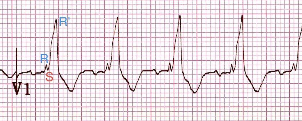
An rsr with widening of the qrs and characteristic findings in other leads is due to a right bundle branch block. 6 mm or S 2mm or rSR with R 10 mm.
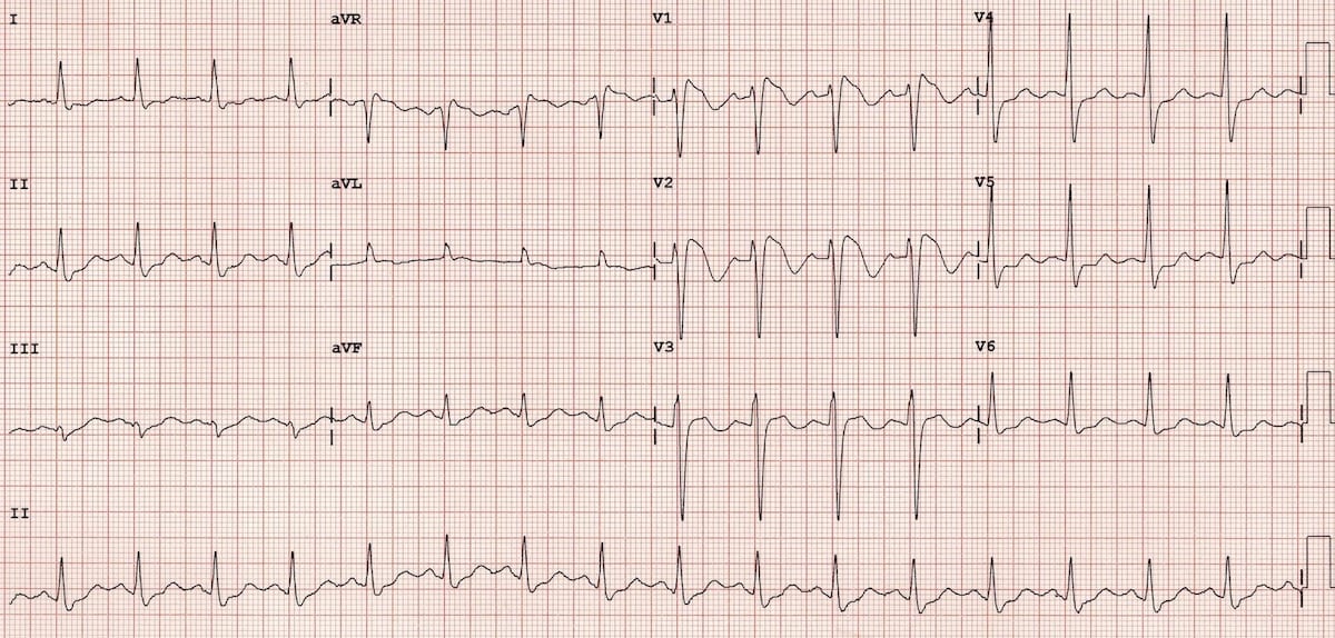
Right Bundle Branch Block Rbbb Litfl Ecg Library Diagnosis
RSR in V1 or V2.
. Newborns have upright T wave in V1. Right ventricular conduction delay means late blood pumping from the right ventricle of the heart. RSR or QR pattern in V1 suggests right ventricular conduction.
It has a characteristic pattern on the ECG with an rSR pattern in the lead V1. Related Questions I might have brugada its only a. Related Questions I might have brugada its only a.
R in V5 or V6 5 mm. 1 doctor answer 3 doctors weighed in. The differential diagnosis of an rSr pattern in leads V1-V2 on electrocardiogram is a frequently encountered entity in clinical cardiology.
One of the more frequent dilemmas in ECG interpretation is the differential diagnosis of an rSr pattern in leads V 1-V 2. Qr in V 1 and the presence of negative T waves in V 2 or V 3 also predict a complicated hospital course and therefore are useful for risk stratification in pulmonary embolism. Normal Sinus rhythm Possible Left Atrial enlargement RSR or QR pattern in V1 suggests right ventricular conduction delay Borderline ECG Anything to worry about.
Goswami Debopom s Response Thank you for posting your query. The right bundle branch taking signals to the right ventricle can often have a conduction delay and the manifestation on ECG is called right bundle branch block RBBB. We often face this finding in asymptomatic and otherwise healthy individuals and the causes may vary from benign nonpathological variants to severe or life-threatening heart diseases such as Brugada syndrome or arrhythmogenic right.
More than 012 seconds. Widened slurred S wave in V6. RS ratio in V5 or V6 1.
T wave remains inverted in V1 throughout early childhood. We often face this finding in asymptomatic and otherwise healthy individuals and the causes may vary from benign nonpathological variants to severe or life-threatening heart diseases such as Brugada syndrome or arrhythmogenic right ventricular. 4 If the QRS is wide the presence of an R in leads V 1 V 2 usually is in the context of a complete right bundle branch block RBBB but other causes have been described.
R in V1 S in V5 or V6 10 mm. ECG results1100 sinus rhythm2420 RSR QR in lead V1V2 consistent with right ventricular conduction delay9130 borderline ECG. It is characterized as a long QRS complex Ie.
An rSr pattern in the right precordial leads is a relatively common electrocardiographic finding that has been described in up to 7 of patients without apparent heart disease. Upright T wave in V1 1 week of age. I can understand your concern.
RSR or QR pattern in V1 suggests right ventricular conduction delay Nonspecific T wave abnormality Abnormal ECG Thu 19 Feb 2015 Report Abuse General Family Physician Dr. Rsr pronounced r s r-prime can be a normal finding in leads v1 and v2. A Verified Doctor answered A US doctor answered Learn more.
One of the more frequent dilemmas in ECG interpretation is the differential diagnosis of an rSr pattern in leads V1 -V2. This finding often presents itself in asymptomatic and healthy individuals. S in V5 or V6 7 mm.
Upright T wave beyond the first week of life is a strong indicator of RVH. RSR in V1 with a tall R 10 mm qR pattern in V1. Right bundle branch block can exist in the absence of any other significant heart disease and may not do much harm by.
Any one of the following in lead V1. Interpretation on ekg says sinus rhythm Low Voltage in precordial leads - RSRV1-non diagnostic - Horizontal axis for age. Other chest lead criteria.
The causes might vary from benign and nonpathological to. 142 QT316 QTcH372 QRSD96 P-QRS-T47-1041. RSR pattern in V1 suggests right bundle branch block RBBB.
Compared with other ECG signs Qr in V 1 is the strongest predictor of right ventricular dysfunction and it is highly associated with troponin leakage and myocardial shear stress. T wave normally becomes inverted during the first week of life. Read Responses 4 Follow.
Ecg results1100 sinus rhythm2420 rsr qr in lead v1v2 consistent with right ventricular conduction delay9130 borderline ecg. RS ratio 1 and negative T wave.

The Rsr Pattern In Leads V1 V2 Algorithm And Differential Diagnosis Sciencedirect

Differential Diagnosis Of Rsr Pattern In Leads V1 V2 Comprehensive Review And Proposed Algorithm Baranchuk 2015 Annals Of Noninvasive Electrocardiology Wiley Online Library

The Rsr Pattern In Leads V1 V2 Algorithm And Differential Diagnosis Sciencedirect
Right Bundle Branch Block Rbbb Litfl Ecg Library Diagnosis

Rsr In V1 Resources

Right Bundle Branch Block Rbbb Litfl Ecg Library Diagnosis

Electrocardiogram Right Axis Deviation Right Ventricle Overload Qr Download Scientific Diagram

Dr Smith S Ecg Blog Rsr With St Elevation Is This Right Bundle Branch Block With Stemi Type 2 Brugada
0 comments
Post a Comment